Brain landscape: anatomy and pathways
Cortex · deep structures · somatotopy · motor and sensory pathways
Frontal cortex
- Prefrontal networks: planning, working memory, judgement and behavioural regulation
- Precentral gyrus: primary motor cortex (M1; area 4)
- Premotor and supplementary motor areas: preparation and sequencing of actions
- Inferior frontal language networks: speech production, usually left-dominant
Parietal cortex
- Postcentral gyrus: primary somatosensory cortex (S1; areas 3, 1, 2)
- Touch, position sense and integration of body signals
- Posterior parietal networks: spatial attention and visually guided action
- Association regions support body schema, praxis and multisensory integration
Temporal and occipital cortex
- Superior temporal cortex: auditory processing and language comprehension networks
- Medial temporal structures: hippocampal memory formation
- Primary visual cortex (V1; area 17): around the calcarine sulcus
- Dorsal visual stream: spatial/action processing; ventral stream: object recognition
Deep structures
- Thalamus: major sensory and motor relay, with reciprocal cortical connections
- Basal ganglia: caudate, putamen and globus pallidus; movement selection and scaling
- Hypothalamus: homeostasis, endocrine and autonomic regulation
- Corpus callosum: communication between hemispheres; internal capsule: projection fibres
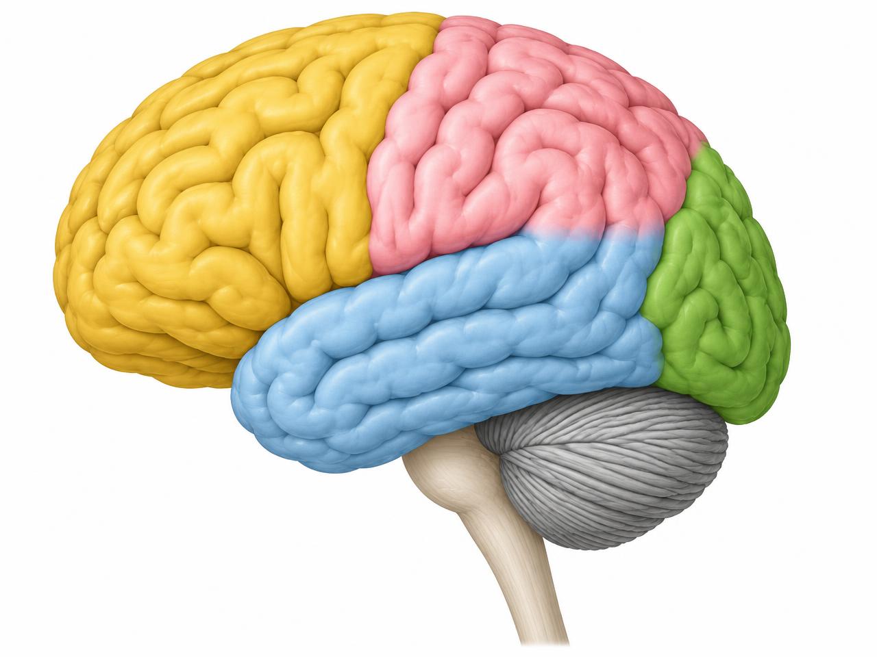 123456
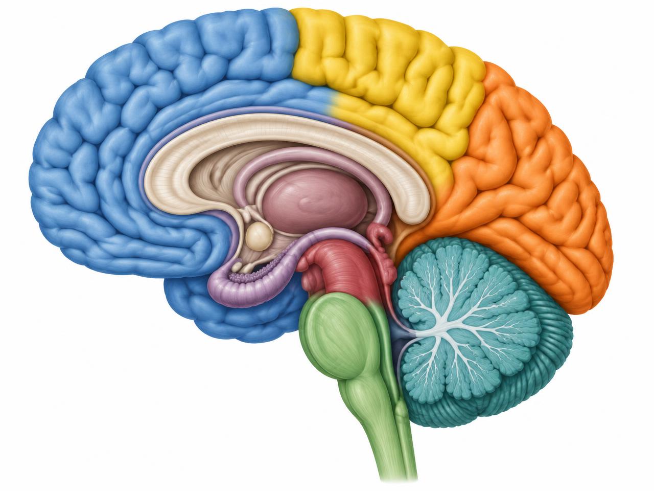
Medial view: corpus callosum, diencephalon, brainstem and cerebellum
1 Frontal lobe2 Parietal lobe3 Temporal lobe4 Occipital lobe5 Cerebellum6 Brainstem
Cortical body representation
Motor map (M1)
- Medial → lateral: leg / foot → trunk → arm / hand → face / tongue
- Leg representation extends onto the medial paracentral lobule
- Hand and face occupy relatively large areas; maps overlap and are adaptable
Sensory map (S1)
- A broadly similar medial-to-lateral body arrangement lies behind the central sulcus
- Area 3a: mainly proprioceptive; 3b: mainly cutaneous; 1 and 2: further tactile/body processing
- Cortical representation reflects sensory resolution, not body-part size
Corticospinal tract
- Motor cortex → corona radiata → internal capsule → cerebral peduncle → pons → medullary pyramids
- Most fibres cross in the caudal medulla, then descend in the lateral corticospinal tract
- Spinal circuits → lower motor neurons → peripheral nerves → skeletal muscle
Dorsal columns / medial lemniscus
- Discriminative touch, vibration and conscious proprioception
- Receptor → dorsal root → ipsilateral dorsal column → medulla (gracile/cuneate nuclei)
- Cross in medulla → medial lemniscus → VPL thalamus → S1
Anterolateral system
- Pain and temperature; also crude touch
- Receptor → dorsal root → dorsal horn → spinal crossing near entry
- Contralateral ascent → thalamic and brainstem targets → cortical networks
Spinal cord in the vertebra and nerve roots
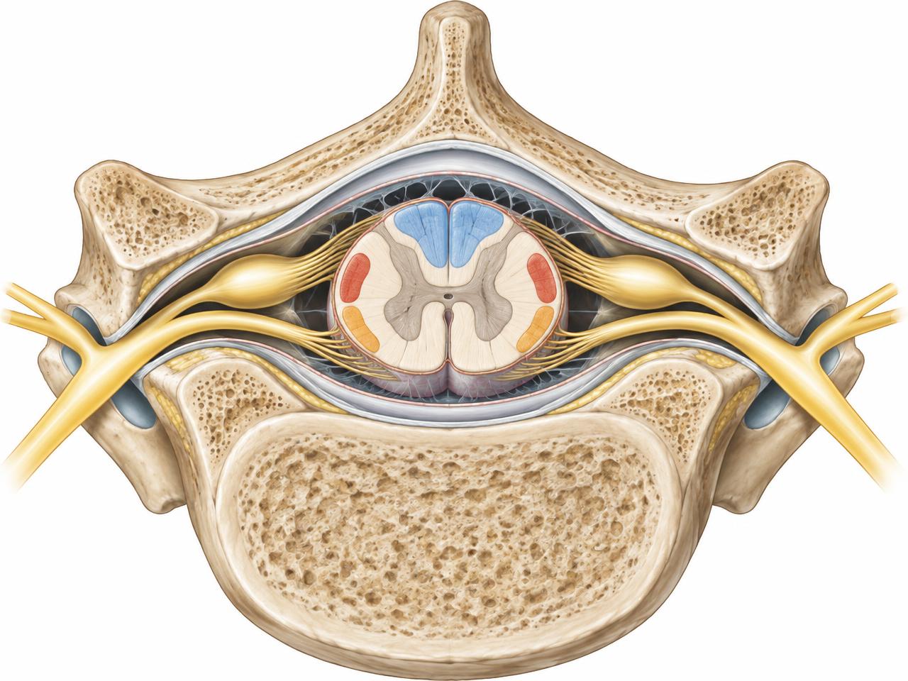
Axial view from above: posterior (spinous process) at top, vertebral body at bottom
Colour key and root anatomy
- Blue — dorsal columns (gracile / cuneate fasciculi): touch, vibration, proprioception → medial lemniscus
- Red — lateral corticospinal tract: descending voluntary motor commands
- Orange — anterolateral (spinothalamic) system: pain and temperature
- Grey matter “butterfly”: dorsal horns (sensory) and ventral horns (lower motor neurons)
- Dorsal root with dorsal root ganglion (sensory cell bodies) + ventral root (motor) → spinal nerve
- Spinal nerve exits through the intervertebral foramen; cord is wrapped by dura, arachnoid and pia mater
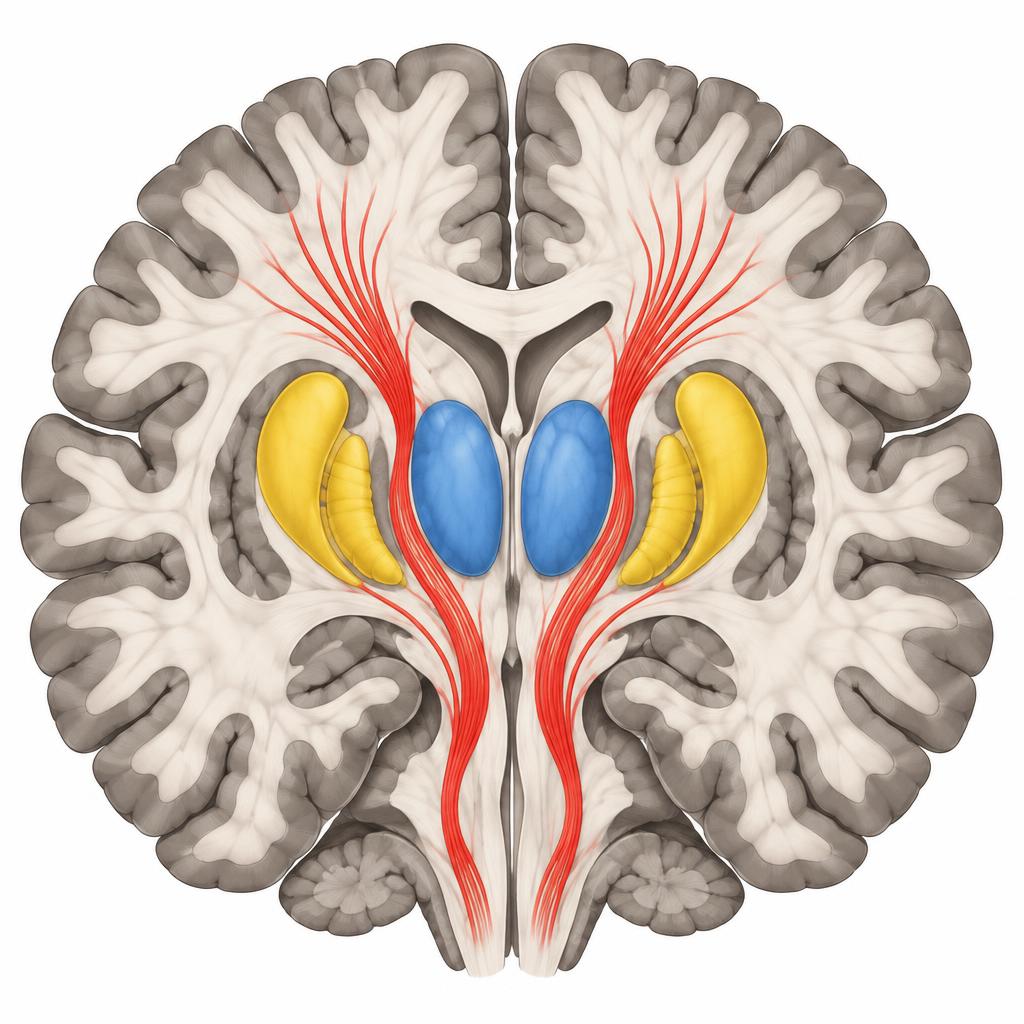
Network interpretation
- Both hemispheres participate in most complex functions; lateralization is not a personality classification
- Cerebellum: coordination, timing, error correction and adaptation
- Clinical localization combines history, strength, reflexes, sensation, coordination and imaging when appropriate
- Illustrative atlas views; simplified maps are not patient-specific diagnostic images